Nature Special Edition
Welcome, everyone! Welcome to this very special edition of Mind the Graph Experience in our blog!
In Mind the Graph Experience Blog articles, you are going to see science figures from published articles created using the platform Mind the Graph.
You can check out the previous editions here.
This is a very special edition. We gather all science figures, graphical abstracts, and infographics produced in the Mind the Graph platform found in Nature articles.
So, if you are submitting your article to Nature and looking for inspiration to create the visual communication part of your manuscript, this article is for you!
Now, without further delay, let’s get into business. See below images and the articles from Nature.
Mind the Graph Illustrations on Nature.
If you are still not a Mind the Graph user, what about create your account right now? Try for free. Click here to Start Now.
Have you ever saw how many illustrations Mind the Graph platform has available for you to use? No? Visit our Gallery. I’m sure you will find out everything you need to create your graphical abstract.
Still, if you didn’t find what you need, we have a solution for that: at Mind the Graph, you can request a personalized illustration for our high expert illustrations team. They can do any illustration you may need.
All you have to do is login into your account at Mind the Graph and fill the Request an Icon form to send us your idea and a reference image. In a week’s tops, you will get your requested illustration inside your workspace.
Sounds amazing, uh? The personalized illustration service is available for subscribers’ users. Check our Mind the Graph subscriptions plans and see what fits best for you.
Now, article number 1 below has a graphical abstract created by the Becker Laboratory for “Metabolic regulation of gene expression by histone lactylation”. Before I give you any more information about the article, see if you can understand the main message of the graphical abstract below.
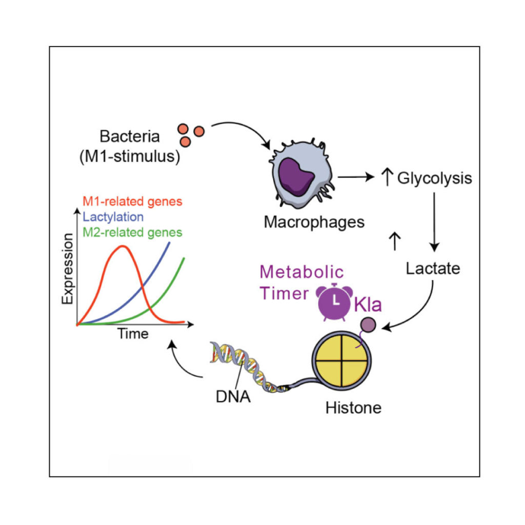
Now, In the article words, the study “results suggest that an endogenous ‘lactate clock’ in bacterially challenged M1 macrophages turns on gene expression to promote homeostasis”.
For more complete and further information, check out the full article here.
Article number 2 has two figures created using Mind the Graph. “Modeling of inhomogeneous electromagnetic fields in the nervous system: a novel paradigm in understanding cell interactions, disease etiology and therapy” was published in August 2018.
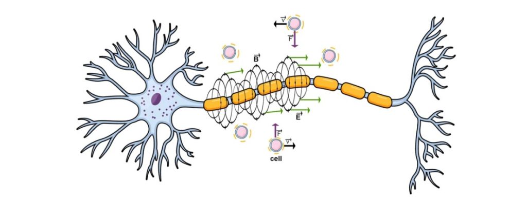
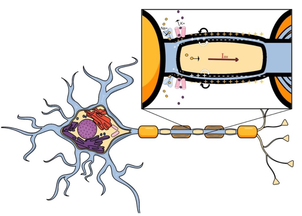
In the words of the article, the study “presents a novel model of inhomogeneous electromagnetic fields originating from nerve fibres and delineates their influence on cells”. The first figure shows the “forces acting upon cells in the vicinity of the neuron,” and the second one shows a “proposed axial and longitudinal current flow generation within a neuron”.
Article number 3 is called “Acellular bioscaffolds redirect cardiac fibroblasts and promote functional tissue repair in rodents and humans with myocardial injury”, published in June 2020.
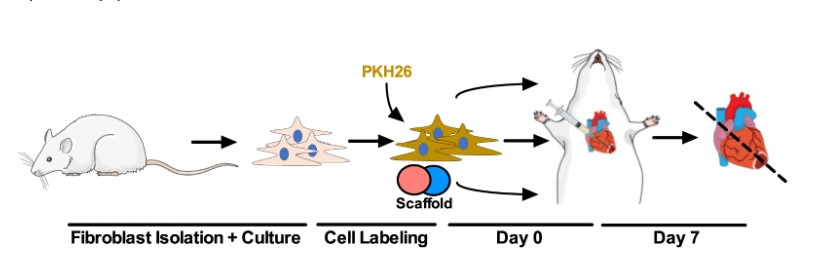
The image above is just a part of a bigger image that shares some information about the methodology, and the full image includes results and graphics. Click here to read the full article.
These three following images are from article number 4, called “Amazon rainforest rodents (Proechimys) are resistant to post-stroke epilepsy”, published in August 2021.
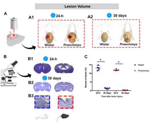
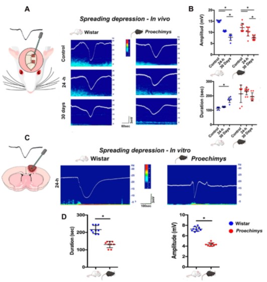
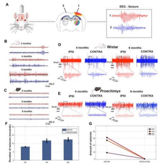
The present study had the main goal to present to compare “ (1) stroke-related brain responses 24-h and 30 days after cortical photothrombosis and (2) post-stroke epilepsy between Proechimys rodents and Wistar rats” being Proechimys, an Amazonian rodent, as resistant to acquired epilepsy development in post-status epilepticus models and Wistar rats, a traditional animal used for laboratory research. The study had interesting results. Click here to read the full version of the article.
The next article, number 5, was published in July 2020 and named “Beneficial effects of IL-4 and IL-6 on rat neonatal target cardiac cells”.
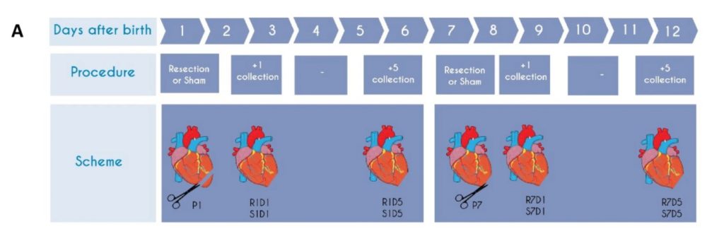
The figure above is only part A of a bigger image. The image shows a part of the methodology performed by the study: “Cardiac apex resection or false surgery was performed in 1-day-old and 7-day-old rats, and samples were collected one (D1) or five (D5) days after the procedure”. To read the full article, click here.
Article number 6, published in August 2018, is called “High-resolution synchrotron-based X-ray microtomography as a tool to unveil the three-dimensional neuronal architecture of the brain”. The article is one of the Top 100 in Neuroscience Collection.
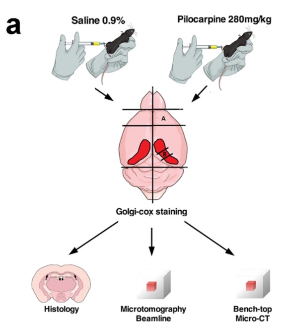
The image is part A, of a bigger picture. The image is a schematic illustration of the experimental procedures, that is, a summary of the adult mice were injected with saline or pilocarpine solution, then the brains were removed, and the frontal cortex and medial part of the hippocampus were dissected. To read the full article, click here.
And that’s all folk!
I hope you enjoyed seeing these science figures and find it helpful to create yours.
Create your science figures doesn’t have to be hard and painful, all you need is to follow a great step-by-step and have amazing visuals references. And since you already have those in there step-by-step, you can find them here.
What about give it a try right now?
Take the first step,
and start now!


Subscribe to our newsletter
Exclusive high quality content about effective visual
communication in science.