
We use anatomical positions to describe the body because they provide a reference point. They can be used similarly to standardization tools to describe an organism’s position. Medical practitioners cannot discuss patients’ bodies without having a general awareness of anatomical position, which is particularly important in medicine.
There is a standard anatomical terminology for each species of organism. In this article, we will learn more about anatomical positions and why they are so important.
Anatomical position: what does it mean, and why is it so important?
The term anatomical position refers to how an organism’s body is oriented, located, moved, and directed. A person standing upright, palms facing forward, thumbs pointed away from their body, with arms hanging by their sides, follows this universal standard of anatomically upright posture. Foot alignment is slightly parallel, with toes pointing forward.
There is no literal meaning to anatomical positions. They are just references for the way a body is placed. As with other anatomical terms, for example, body planes, directions, and relationships, this word must be viewed in context.
The anatomical position is essential for understanding human anatomy and physiology because it provides a precise and uniform way to describe these body structures. Individual anatomical positions are described using various anatomical terms describing their relative placement to standardized positions.
For instance, posterior or dorsal refers to your back, and proximal and distal refer to the distance between you and something. Whenever anatomical terms are used in a standard manner, they become clear.
Anatomical position jargon: what it means
There are many different terminologies used to describe anatomical positions. In the absence of a clear understanding, these can be confusing. Firstly, let’s take a look at the terminologies and then describe the main types of anatomical positions.
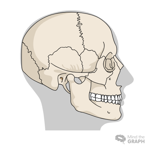
Regions of the body
Five areas make up the human body: the head, the neck, the torso, the upper limbs, and the lower extremities.
The skull and face are part of the head region. The chest, abdomen, and pelvis comprise the torso or trunk. Shoulders, armpits, and arms are considered upper limbs. A lower extremity consists of the legs, buttocks, thighs, knees, ankles, and feet.
This division facilitates identifying and categorizing specific body parts.
Anatomical planes
Various organs and body structures are cut or sliced by these imaginary planes that intersect the body. Body planes are divided into three categories:
1. The sagittal plane cuts the body longitudinally along its midline. It is parallel to the midline and slices it longitudinally into the left and right halves.
2. The body’s anterior and posterior sections are divided by a vertical line known as the coronal plane.
3. In a transverse plane, the body is divided into superior (upper) and inferior (lower) lobes at right angles to the sagittal and frontal planes.
Directional terms
These adjectives describe the relative placement of two structures within the anatomical space. The directional terms include:
- Anterior: Frontal view of the body (at or near the front)
- Posterior: Backside of the body (back view)
- Midline: An imaginary vertical line dividing the body evenly (at its center).
- Lateral: Away from the midline (side view)
- Medial: Closer to the midline (side view)
- Superior: Looking up at a structure (bird’s-eye view)
- Inferior: Looking up from the bottom of a structure (away from the head/lower part)
- Superficial: Located close to the body’s surface
- Deep: Far from the body’s surface
- Proximal: Closer to a structure’s origin
- Distal: Further away from the point of origin
- Cranial: Headward
- Caudal: Tailward
Cavities
The body is filled with open fluid-filled spaces called cavities, which enclose different types of anatomical structures. These are located in axial locations, including the skull, vertebral column, thorax, and abdominal cavity.
Cavities compartmentalize the body, protect organs, and lubricate them, reducing friction while moving the organs. There are two main cavities in the body: the ventral cavity and the dorsal cavity.
The Ventral cavity is a large cavity partitioned by a dome-shaped respiratory muscle called the diaphragm into the thoracic cavity and abdominopelvic cavity. The heart, lungs, trachea, esophagus, large blood vessels, and nerves are all located in the upper ventral cavity or chest cavity.
The Lower ventral cavity (abdominopelvic) comprises the abdomen and pelvis. In the abdominal cavity are the kidneys, adrenal glands, and most of the gastrointestinal tract. There is a large amount of urogenital system and rectum in the pelvic cavity. There are three boundaries to the pelvic cavity: the abdominal cavity, the sacrum, and the pelvis.
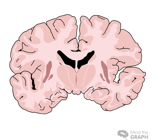
The dorsal cavity is the smaller of the two main cavities. Named for its location in the body, it contains organs that are located more posteriorly. There are two parts to the dorsal cavity. Lower portions, or vertebral canals, house the spinal cord, and the upper portion, or cranial cavity, contains the brain.
Anatomical Positions
Anatomically, there are four main positions: supine, prone, right lateral recumbent, and left lateral recumbent. It is important to note that each position has its medical implications.
- The supine position requires the face and upper body to be turned upwards in a horizontal position. The ventral side of the body is up in supine position, while the dorsal side is down.
- The prone position involves lying flat on the back with the head and upper body looking down. The dorsal side of the body is up in the prone position, and the ventral part is down.
- Right lateral recumbent refers to lying on one’s right side. A patient’s left side is easier to access from this position.
- Compared to the right lateral recumbent position, the left lateral recumbent position is the opposite. The individual is lying on his or her left side in this position. Right-side access is easier from this position.
Communicate your ideas and findings with no effort through impactful visuals
When anatomical positions are illustrated well, understanding them becomes much easier. The use of impactful visuals can facilitate an understanding of even the most difficult or complex research. Read our blog “Scientific Illustration: The key to a world of visual science“.
With Mind the Graph you can illustrate any research and also spread your work to a wide audience. Our library has a large collection of illustrations that you can choose from for your projects. As well as this, you can also customize it from our expert team to meet your specific needs. Visit our website to learn more.
Additionally, errors are made in scientific figures because of a low degree of consistency in color, pattern, and format, incoherence between concepts and designs, disregard for discipline-specific codes, inconsistent proportions and angles, the use of difficult-to-understand words and symbols, and the absence of graphs with accurate scales.


Subscribe to our newsletter
Exclusive high quality content about effective visual
communication in science.