To understand how a neuron works there are two concepts that we need to take a look at. The first one is related to what happens inside the neuron when the information is passing forward – the action potential – the second one is how an information jumps from one to another cell – the synapse. With these two processes, cells of the nerve system are capable to carry on the most complex information throughout the whole body moving it neuron to neuron until finally reach the target cell. In this article, we are going to discuss the first concept, the action potential.
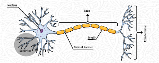
The nerve impulse is an electrochemical signal; it is the main mechanism used to carry information inside a neuron. The dendrites of some neuron detect and receives the impulse from a previous cell, the nerve impulse travels along going from the dendrites to the nucleus than to the axon, and finally to the axon terminal when the impulse is passed to the next neuron. This process goes on repeatedly until it reaches the target cell.
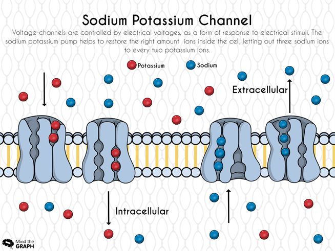
The electrochemical signal is generated due to the movement of ions between the inside and the outside part of the neuron plasma membrane. The ions go from outside to inside, producing a difference of potential in the membrane. The “bridge” used by these ions to go inside the cells is a transmembrane protein called voltage-gated ion channels.
These voltage-channels are controlled by electrical voltages, as a form of response to electrical stimuli, in another words, these channels are not always open to the passage of ions, they only open and close over some electrical voltage stimuli.
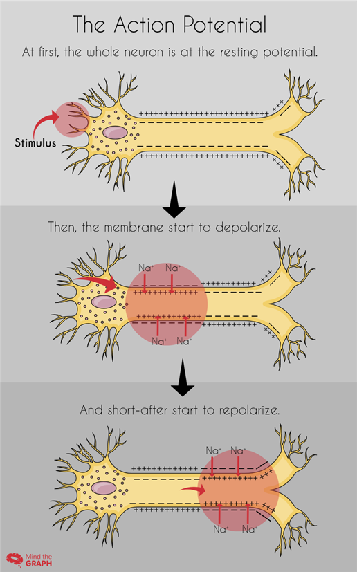
When the cell is not under stimuli, when the membrane is at rest, a potential difference is maintained between the inside and the outside part of a neuron. At rest, the membrane has a potential of -70mV, a negative potential, while the outside have a positive potential. This potential difference is called resting membrane potential, and it is maintained mostly by sodium and potassium ions through the sodium potassium pump.
Under electrical voltage stimuli, the membrane potential difference starts to invert itself, sodium channels open allowing many sodium ions to go inside the cell, turning the membrane momentarily depolarized, or better, the sodium ions turn the inside region of the membrane into a positive net. This movement of depolarization is the famous action potential; the membrane potential rapidly rises and falls. The potential rises to +40mV in a little more than 2miliseconds and go back to the resting state in less than 3miliseconds.
The action potential doesn’t happen in the whole neuron at once, the depolarization of the membrane starts in the dendrites and then to the nucleus part-by-part, depolarizing and going back to the resting state potential shortly after.
To restore the resting membrane potential, the sodium channels close and voltage-gated potassium channels open, allowing potassium ions to go inside the cell, repolarizing the membrane, turning the inside region of the membrane negatively charged again, and the outside region positive. The sodium potassium pump helps to restore the right amount of each ion inside the cell, letting out three sodium ions to every two potassium ions.
We can imagine it like a synchronized movement, from the moment of action potential response to the moment of restoring the resting state.
Interestingly, while in the axon we have the voltage-gated ions channels generating and propagating the nerve impulse, in the dendrites these channels do not exist. In these neuron regions, the signal is passed not by the action potential but a graded potential, a different form of signal propagation, in which the signal scale increase along the way, until turn into the action potential on the axon.
Note that the sodium ion is the one responsible to propagate de action potential, and the potassium to reestablish the resting state. The lack of these ions in the organism can cause problems in quality and efficiency of the action potential, meaning problems at synapses and in the passage of information through the nerve system. All these problems can trigger mental health complications and illnesses.
Coming up, the next step would be the passage of the nerve impulse to the next neuron. A different thing happens in the space between the two neurons, in the synaptic cleft. The synaptic cleft is a very important place to look at and to study, it is where many different neurotransmitters come into action, activating a new signaling pathway using receptors, other proteins and ions besides sodium and potassium. But that we will leave to the next discussion in the article Nerve Impulse PART 2 – The synaptic cleft.
Did you liked the infographs in this article? You can use Mind the Graph and made pictures informative like this too. Get at Mind the Graph and take a look of the illustration gallery, there available neurology and biochemistry illustrations, and if you need any help please contact us!


Subscribe to our newsletter
Exclusive high quality content about effective visual
communication in science.